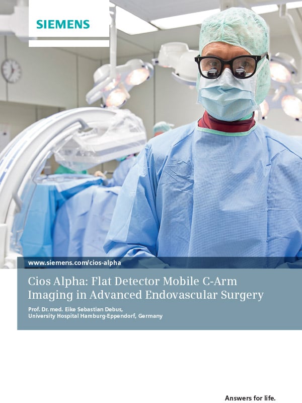
Improving TEVAR & Vascular Surgery with High-performance Intraoperative Imaging
Case Study
A 250-pound, 54-year-old man suffered a type B aortic dissection in the wake of a motorcycle accident, which had become chronic by the time the intervention was performed. The large false lumen located directly below the left subclavian artery exit prompted the surgery team to perform a thoracic endovascular aortic repair (TEVAR).
This procedure is not new for Professor Dr. med E. Sebastian Debus, director of the Clinic for Vascular Medicine at University Hospital in Hamburg, Germany.
One thing is different this time: Professor Debus is using the Siemens Cios Alpha flat detector mobile C-arm.
Complete the form below to read the case study.

Download this case study to learn how the Cios Alpha improves vascular surgery, such as:
- Flat detector replaces round image intensifier
- See more of the surrounding structures, in addition to the relevant section of the aorta, with a large field of view (FOV)
- Observe how contrast agents disperse in the renal arteries branching off from the aorta in the same FOV
- Permits carbon dioxide to be used for patients unable to tolerate contrast agents
- Powerful tool to overcome obesity and acquire high quality images
Click here to learn about our innovative C-arm technology, or see our complete surgical solutions.
About Cassling
Cassling, founded by Bob Cassling in 1984, is an Advanced Partner with Siemens Healthineers. As a family-owned company, we're dedicated to strengthening healthcare in local communities.
Copyright © 2023 Cassling. All Rights Reserved. Compliance Policy. Privacy Policy.