Strengthening Cardiac Care Through Innovative Technology
Case Study
Kansas Heart Hospital Improves Patient Outcomes with 3D Transesophageal Echocardiography
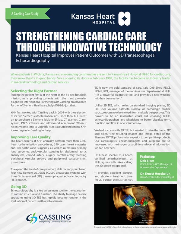
Echocardiography is a key assessment tool for the evaluation of cardiac structure and function. The ability to image cardiac structures using 3D transesophageal echocardiography
(TEE) has rapidly become routine in the evaluation of patients with a valve disease.
The heart experts at Kansas Heart Hospital, located in Wichita, Kansas, annually perform more than 2,500 heart catheterization procedures, 350 open heart surgeries and 100 aortic valve surgeries, plus many more advanced heart procedures.
Download our case study to learn how KHH expanded their level of care and improved patient outcomes with Siemens ACUSON SC2000 ultrasound systems and 3D TEE probes.
Fill out the form below to access the case study.
In Their Own Words:
"The 3D technology allows us to definitively answer questions that could only be surmised under 2D."
-Dr. Ernest Hoeckel, Jr.
Anesthesiologist, Kansas Heart Hospital
“The 3D TEE has been a huge success at Kansas Heart; it’s amazing what we can accomplish in real time.”
-Deb Sikes, RDCS, RDMS, RVT, Manager of Non-Invasive Department, Kansas Heart Hospital
