New Ultrasound technology in detecting liver lesions, ultrasound’s role in the fight against COVID-19, and more…
Are the days of invasive tests behind us? Not yet. But ultrasound is bringing us closer than ever before, and liver imaging provides a good example.
Contrast-enhanced ultrasound
Advances in ultrasound, known for its safety, low cost, patient tolerability and system mobility, are especially promising. The most recent is contrast-enhanced ultrasound (CEUS), an emerging technology used in cardiac and abdominal imaging for decades outside the U.S.
The introduction in the 1980s of ultrasound contrast agents utilizing microbubbles paved the way for modern CEUS. With their high molecular weight core and surrounding lipid or albumin shells, these microbubbles allow for longer imaging times.
One of contrast-enhanced ultrasound's most important attributes is the ability to obtain real-time imaging, compared to contrast-enhanced CT or MRI, which provide only single 'snapshots' in time that could result in a missed diagnosis.
Since the Food and Drug Administration approved it in 2016, CEUS has impacted the diagnosis of liver lesions. It's also made an impact on imaging providers and patients.
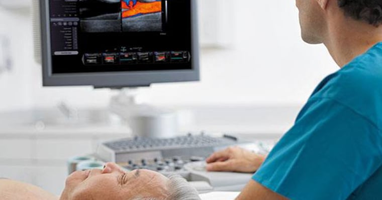
In the past, lesions like these would probably have required a biopsy to identify. CEUS now provides a non-invasive option that captures an accurate image at three separate stages to diagnose the type of lesion affecting the patient. It's radiation-free, safe, and in many settings, a highly sensitive imaging modality.
The reduction of invasive procedures coupled with a dramatic rise in non-invasive ultrasound can lead to faster results, less risk of infection and increased patient and provider satisfaction.

New data suggest CEUS has potential as a diagnostic imaging tool in cases where CT or MRI are not viable options for patients. It also has potential for diagnostic imaging of other conditions and diseases, such as kidney lesions. Since approving ultrasound imaging for liver lesions, the FDA has given the green light to additional applications of contrast-enhanced ultrasound.
The Role of Ultrasound in COVID-19
COVID-19 continues to place heavy demands on healthcare workers in 2021, as they struggle with increasing numbers of critically ill patients, and variants continue to emerge. But ultrasound has made a substantial contribution in fighting the pandemic.
COVID-19 patients often endure constantly changing conditions requiring ultrasound results to examine the lung and pleural space. Although high resolution computed tomography (HR-CT) is the gold standard for thoracic evaluation, moving COVID patients from the Intensive Care Unit (ICU) for this diagnostic imaging can be problematic, even harmful. And while chest X-ray (CXR) is considered the standard of care for many diagnostic tests in the ICU, it can be a limited option for patients who may have the highly contagious SARS-CoV-2.
Lung ultrasonography provides results similar to HR-CT. It's also superior to standard CXR in evaluating pneumonia. It provides a lower-cost option, is easier to use at point of care, non-invasive and doesn't emit radiation.
And as far as imaging systems go, it's easy to maintain the cleanliness of ultrasound systems if you use the list of appropriate disinfectants and procedures provided by the manufacturer.

Comfort follows form and function in system design
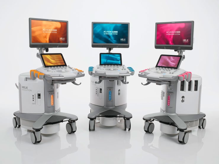
The newest ultrasound systems can be customized to your preferences when performing an exam. They also pack in all the technologies you've come to rely on, and then some.
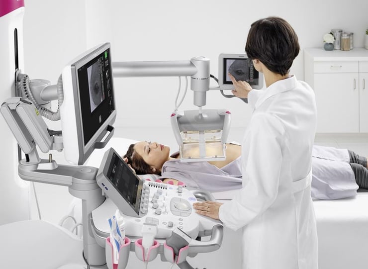
System ergonomics continue to evolve and consider user comfort. With large, adjustable LED screens and the ability to configure the height of the monitor, angle of the control console and placement of the machine to your preferment, you'll experience less strain on your body than before. So, while the patient benefits from new and improved imaging capabilities, you'll benefit from ergonomic improvements.

What's important to note is that all of this happens without any loss of diagnostic capabilities. Far from it.
High-contrast resolution, high-fidelity transmission, one-touch image optimization, improved elasticity, stunning image detail and automation continue to resonate in ultrasound. Hospitals and clinics continue to find new ways to improve clinical workflows and patient-to-provider relationships.
Department to Department Mobility
The increased versatility of the modern ultrasound systems means you can wheel virtually anywhere – perfect for mobile ultrasound units that travel around the hospital.
Less space means less weight and more opportunity for comfortable placement. Wheel the unit next to the patient's bedside in virtually any hospital room. (That's especially important now, with COVID patients who need to remain quarantined in a single space.)
The system can also remain stationary to create an ultrasound suite out of almost any available space in your facility.

The adaptability of newer ultrasound systems makes switching between different types of patients and their necessary exams easier than ever.
Readily reproducible workflows have always been the goal. With machine-assisted guidance, it's possible to conduct more exams more quickly without ever losing time or image accuracy. Workflow efficiency, revenue, and the patient experience could all improve dramatically in the coming years artificial intelligence applications evolve the imaging field in unprecedented ways.


Comments