
 Chronic liver disease (CLD) and cirrhosis are major health problems in the United States. In 2018, the Centers for Disease Control reported that 4.5 million adults were diagnosed with liver disease. In 2019, CLD and cirrhosis claimed more than 44,000 American lives. And according to a 2018 study by Intermountain Medical Center, nonalcoholic fatty liver disease alone cost the U.S. healthcare system $32 billion annually.
Historically, biopsy has served as the gold standard for clinical assessment of liver disease. But it’s invasive, expensive and can be painful. It’s not an ideal method of repeated assessment of a disease’s progression. The healthcare and economic challenges highlight the need for noninvasive and cost-effective technology to diagnose and screen for liver disease.
The good news? There is ultrasound technology that can meet those challenges.
As chronic liver disease is a major problem in the general population, there is a need to diagnose and screen for liver disease with using noninvasive, cost-effective and sensitive techniques.
Elastography is an umbrella term describing technology that uses force displacement to measure and visualize tissue elasticity or stiffness in a region of interest (ROI) and can be used to investigate disease in the liver. Stiffness is usually indicative of fibrosis or steatosis (fatty liver disease), which can indicate numerous disease conditions, including cirrhosis and hepatitis. Elastography is especially advantageous when fibrosis is spread out in clumps.
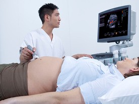
Shear wave elastography (SWE) uses fast-moving shear waves to measure tissue stiffness and provide clinically useful information in real-time and is especially effective in obese patients. Obtaining information quickly, accurately, and without discomfort can generate benefits to the patient as well as the practice.
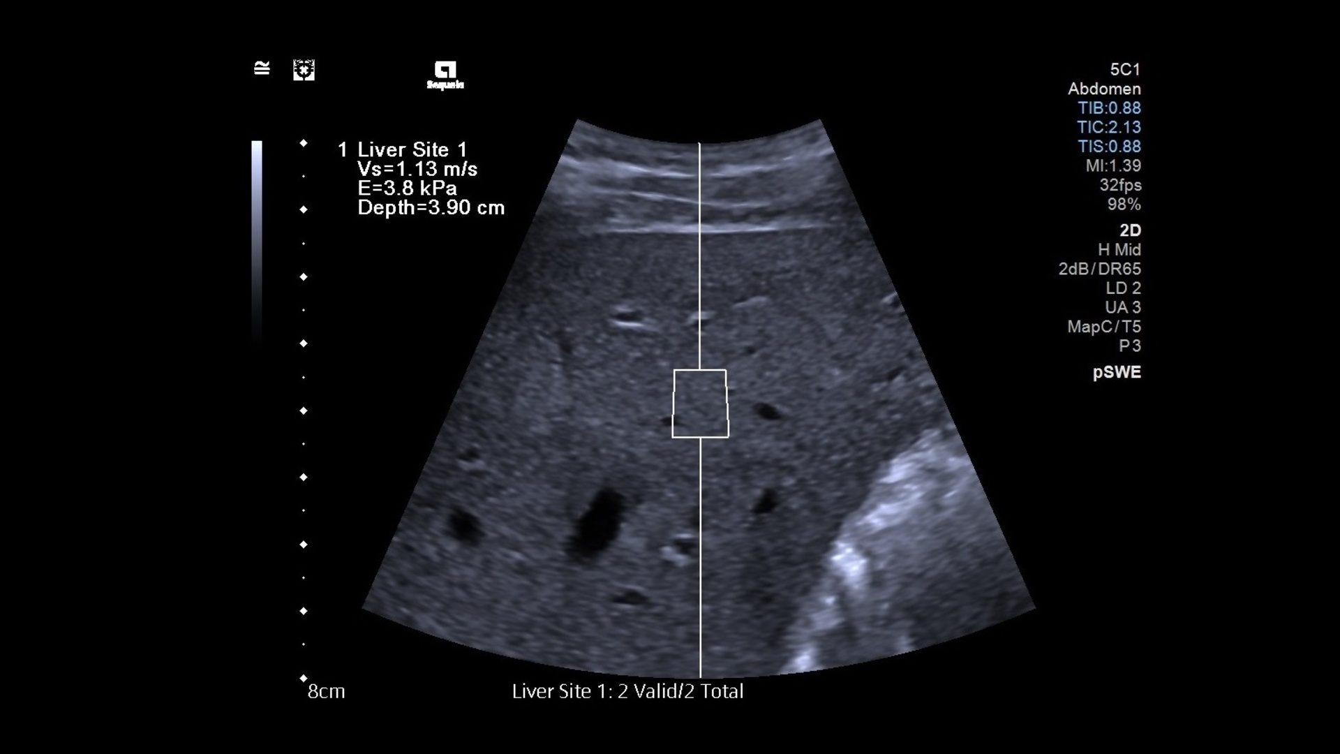
In SWE, an acoustic radiation force (a ‘push’ or pulse) produces shear waves deep in an ROI that travel sideways through the tissue. How fast the waves travel indicates the tissue’s stiffness.
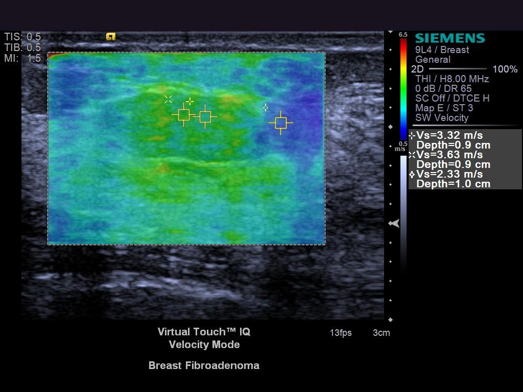
This is translated into a visual elasticity map. Images can be seen in real-time using a normal B-mode ultrasound probe.
SWE is useful in evaluating the musculoskeletal system, and tissue in many different organs, including lymph nodes, prostrate, thyroid, pancreas, breast, spleen and, especially, the liver. It can provide additional diagnostic information than an anatomical image and can be used to guide biopsies. Noninvasive, it could even replace invasive and painful biopsies, entirely.
To read about additional trends in ultrasound, be sure to read part one of Trends in Ultrasound 2022!


Comments