With so many technological advancements happening across all of healthcare, it’s easy to lose sight of the incredible evolution happening to one of the cornerstones of the hospital imaging experience: MRI.
2025 will continue to see a number of improvements to the MRI patient experience, the workflows of imaging teams and the systems that are crucial to ensuring the image is of the highest quality necessary to aid in a successful diagnosis.
Let’s review some of the latest developments in MRI technology to see what will be shaping the conversation around magnetic resonance imaging for years to come.
.55T MRI is Here to Stay
For years, the conventional wisdom held that, in order to get a high-quality MRI exam, you need a field strength of 1.5T or above.
However, that conventional wisdom has been proven wrong with the advent of .55T MRI. Known as High-V MRI, systems with this field strength take the power of digitalization and deliberately apply it to a new field strength of .55T with inherent clinical benefits.
High-V MRI does this in a few different ways:
- Improved implant imaging — Imaging metal implants has historically been difficult with conventional MRI systems because of the artifacts that result. High-V MRI offers intrinsic physical advantages that result in reduced metal distortions, leading to strongly improved diagnostic capabilities for implant imaging.
- Reduced susceptibility challenges — Susceptibility artifacts are a well-known phenomenon in MRI. One prominent example is an air-tissue interface that occurs at the sinuses and orbits. The unique field strength of High-V MRI offers physical advantages that inherently reduce these susceptibility artifacts. This leads to reduced geometric distortions in diffusion imaging, resulting in improved diagnostic quality.
- Pulmonary imaging opportunities — Pulmonary imaging has been notoriously difficult with conventional MRI as the air-tissue interfaces result in a fast signal decay. These challenges scale with magnetic field strength, which makes High-V MRI the perfect opportunity for pulmonary imaging. High-V MRI provides possibilities to expand the reach of your pulmonary MRI capabilities.
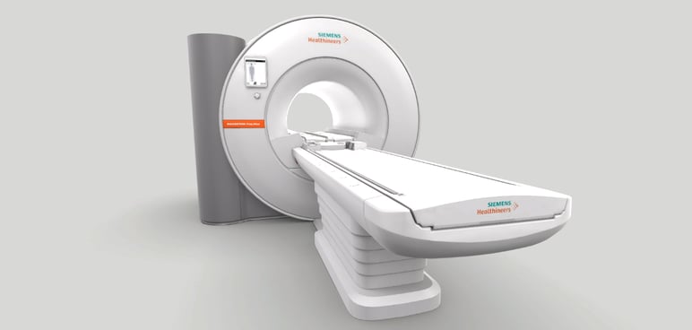
There’s more to explore when it comes to High-V MRI. Start here to learn about how systems like the MAGNETOM Free.Max are causing experts to rethink their ideas about traditional field strength.
Reduced Helium Means New Frontiers in Feasibility (and Sustainability)
Installing an MRI system in a small space is a big challenge; doing it economically is an even bigger one.
In the past, the lack of feasible areas within a hospital complex may have led imaging teams to rely on traveling mobile MRI solutions. Similarly, the complexity of construction and installation of a larger system may have caused departments to stick with outdated systems much longer than they would have preferred.
But recently, the advent of helium-free infrastructure has opened up opportunities for organizations to install smaller, lighter MRI systems in areas where bulkier technology simply would not have been feasible. The less intensive installation requirements of helium-independent systems like the MAGNETOM Free.Max simplify site selection and open access to MRI.
By limiting the presence of helium, numerous advantages become apparent. First, hospitals can make progress toward their sustainability efforts, as helium is a valuable resource that organizations around the world are looking to conserve. Installation becomes easier because there’s no need to worry about a long quench pipe or “helium chimney.” And because you’re saving on the size of the system as well, extensive construction may not be necessary.
Artificial Intelligence
Inroads have been made by artificial intelligence applications across healthcare, and MRI is certainly no exception. AI-powered workflows are here, and they have the potential to totally reshape clinical processes for the better.
Deep Resolve is a fantastic example of the growing advances in AI technologies, whose deep neural networks need to be trained on large datasets to deliver excellent image quality and provide high levels of acceleration to imaging teams.
Launched in 2020 with the introduction of Deep Resolve Gain & Deep Resolve Sharp, these were among the first, most exciting technologies impacting MRI.
Deep Resolve Gain is a targeted reconstruction method to increase signal-to-noise ratio (SNR) of images using the iterative reconstruction process, enabling either shorter scan times or higher resolution imaging based on the needs of the customer. Meanwhile, Deep Resolve Sharp leverages a deep neural network solution that improves the MR data quality by increasing the image resolution.
In 2022, these promising technologies were joined by the latest additions to the Deep Resolve family.
Deep Resolve Boost is an AI-based deep neural network solution leveraging (K-space) raw data-to-image deep learning reconstruction technology that enables high SNR as well as a new level of image acceleration.
Deep Resolve Swift Brain is an MR brain acquisition that employs a multi-shot, echo planar imaging (EPI) approach that achieves all five relevant contrasts for a total brain exam in two minutes. It leverages both a deep learning reconstruction as well as a new static field correction for a fast neuro evaluation when time is critical.
In addition to the Deep Resolve updates, Siemens AI-Rad Companion has introduced two new extensions into the MR space:
AI-Rad Companion Brain MR is a post-processing image analysis software that assists clinicians in viewing, analyzing and evaluating MR brain images as a diagnostic aid, saving radiologists’ valuable time by automatically segmenting, measuring volumes and highlighting over 30 different regions of the brain.
AI-Rad Companion Prostate MR is a cloud-based image processing software that provides quantitative and qualitative information based on prostate MR DICOM images, driving productivity by contouring the prostate gland on the MR image series, while still permitting manual contouring manipulation and annotation of the prostate.
Taken together, Deep Resolve and the AI-Rad Companion solutions are intended to increase patient comfort, decrease image acquisition times while increasing quality and quantitation, and improve staff satisfaction while driving referrals.
Larger Bores
For many reasons, including the reduced amount of helium, it’s now possible to have a larger bore than has been possible in the past.
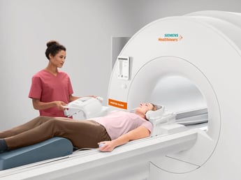
An 80-centimeter bore is a game changer for bariatric patients and others, such as children or patients with claustrophobia, who may have anxiety about their imaging experience. The bigger bore size opens up the possibilities of your imaging workflow, potentially enabling you to receive more patient referrals than would otherwise be possible.
Better access means more opportunities for you and your team.

In-System Entertainment
You can also alleviate patient anxiety by giving your patients the ability to focus on something beyond the exam itself.
In-bore infotainment makes it possible to help a patient get through an exam by providing a high-quality video experience that can be enriched with customized content. The right entertainment option can potentially provide clear audio signals and outstanding sound quality thanks to the cancellation of some of the noise of the MRI itself.
A patient can become immersed in a sound and video experience from the moment they lie on the table, all while being able to hear voice commands from the technologist.
This is truly the next step in enhancing the patient experience. We’re finally seeing the types of technology that would have seemed unimaginable just a few years ago, and it’s going to have a positive impact for years to come. It wouldn’t be a surprise to see this kind of patient-centric tech become standard in the near future.
Enhancements to Liver Imaging
Another new type of technology that’s debuted recently marks an exciting evolution in the liver imaging space.
Because of the required breath holds, liver imaging can be somewhat difficult in comparison to other types of exams. That’s what FREEZEit seeks to solve. By enabling you to overcome the motions inherent to certain types of liver exams, the hope is to aid in the acquisition of clinically valuable images.
StarVIBE delivers robust, free-breathing and contrast-enhanced exams for patients by intelligently resisting motion artifacts. TWIST-VIBE, meanwhile, offers high temporal and spatial resolution with full 4D coverage for multi-arterial imaging with 100% precise contrast timing.
Finally, LiverLab provides non-invasive fat and iron evaluation and enables trending and/or monitoring of patients suspected of having liver diseases, especially in early disease stages. And it’s robust and fast enough to be implemented in routine clinical imaging with only a few clicks.
Be sure to explore these and more if the challenges of liver imaging are something you’re looking to overcome in the near future.
Calculate Your Value
Finally, one thing that’s also new about MRI is the ability to evaluate your potential investment before you commit.
This can be done a couple different ways. First is to use an interactive value calculator to determine the additional revenue your imaging practice may be able to generate with the purchase of a system. You can use the accompanying personalized value brief to determine the financial return to expect. While it’s only an estimate, this level of detail can help you determine when and how an investment in a new MRI system would be right for you.
Another way would be to look at a profit analysis laying out a financial comparison of a representative sample. In this business case, you can determine, for instance, how a mobile MRI solution compares to investing in an in-house system.
There’s a lot of exciting things happening in MRI. Thank you for exploring the ins and outs of the wide variety of MRI systems available to you!


Comments