
Both traditional computed tomography (CT) scanners and cone-beam CT (CBCT) scanners produce pictures of internal body structures. The x-rays use radiation from a radioactive contrast injected into the body to make cross-sectional images.
While both scanners undertake the same primary function, there are technical differences and outcomes that influence whether a standard CT or CBCT would be the best choice for a particular procedure, such as a lung tissue biopsy in interventional pulmonology.
A conventional CT utilizes a rotating, high-output anode X-ray tube and records data with a fan-shaped beam onto image detectors placed in an arc around the patient. It produces a single image per scan. The slices must be overlapped to create a good picture of the area of interest, and the patient must hold their breath throughout the scanning procedure to produce a high-quality image.
A CBCT scanner uses a cone-shaped beam radiating from an X-ray source that covers a large volume with a single rotation. Algorithms construct real-time, high-resolution images from the X-ray images.
Accuracy and Precision: When cone beam technology changes the game
Although cone beam CTs are gaining ground, most hospitals and imaging centers still use a conventional CT scanner. But there are instances in which CBCT technology is a game-changer, especially for interventional pulmonology procedures like CT-guided lung tissue needle biopsies that require placement confirmation – in real-time.
The lung is dynamic, moving constantly. This is a challenge for interventional pulmonologists and other specialists in conducting biopsies, especially when the nodules are deep within the lungs. This is where CT guidance plays an important role.
Until recently, interventional pulmonologists or surgeons have relied on CT images to guide the placement of a biopsy needle. But the images used were static; they might have been taken days, even months before the biopsy. The dynamic action of the lungs meant the nodule might not be precisely where the image indicated, and the result could be a biopsy with lower precision and accuracy.
Biopsies complemented by systems with cone beam technology allow the surgeon or specialist to confirm the placement of the needle within the nodule in real-time. And improving the rate of tool-in-lesion confirmation is the strongest predictor of biopsy success and higher diagnostic yield.
The benefits of cone beam technology in guided procedures extend beyond the patient to the hospital or facility. New technologies, for example, lung cancer therapies, are more easily introduced. It attracts patients and increases revenue. Adding CBCT to a facility creates a concentrated procedural practice, expanding the workforce in other areas of need and enhancing market penetration.
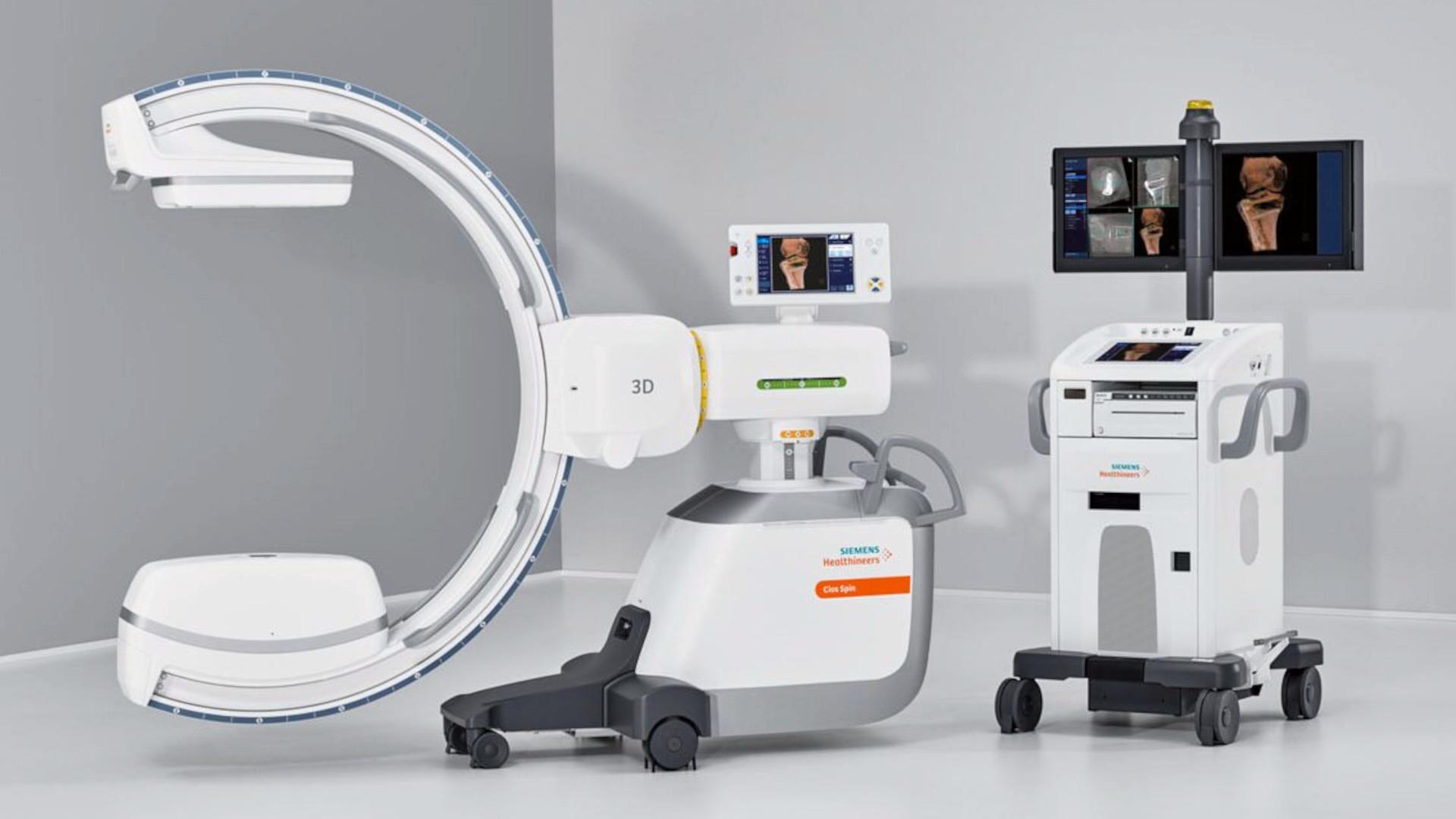
If you’d like to learn more about CBCT technology in systems like the Cios Spin, and how they can improve patient outcomes, call or email your Cassling account representative.


Comments