
When I’m talking with staff at ambulatory surgery centers or hospital surgical departments, one question I typically get is about the technology at the center of the C-arm system:
“Should we go with a flat panel detector or an image intensifier?”
While that might sound like a simple question on the surface, the answer is somewhat complicated once you dig a little deeper. The fact is, there’s no one-size-fits-all solution that can be applied to every facility. Every office-based lab, clinic and healthcare system has very specific needs that must be taken into account.
For this reason, I like to have an in-depth conversation with surgeons and the administrative team to really understand their challenges and what they’re looking to achieve as they pursue new equipment options. Armed with the right information, I can make a recommendation as to the technology that would give an optimum return on investment, plus I can steer them toward a purchasing plan that makes the most sense, i.e. a rent-to-own program versus a fleet swap or a capital investment.
From system features to equipment size, there are strengths for each of these technologies. If you’re in the market for a mobile surgical C-arm, I hope the following information will help inform your research and your purchase decision as you look to expand the size or scope of your practice.
Dose
One of the most significant differences when it comes to flat panel detectors and image intensifiers is the radiation dose.
While the technology powering both types of systems has progressed dramatically over the years, with each new iteration designed to reduce the amount of radiation the patient and the healthcare team may be exposed to, there is a limit to what an image intensifier can accomplish. The simple fact is that flat panel technology will almost always give you a lower dose than an image intensifier.
You can really see this come into play when working with the magnification modes available on each system. In order to get down to the Zoom 3 setting on an image intensifier, you’ll need to increase the dose by five times as much as you would in the normal full field mode. The same magnification on a flat panel detector, on the other hand, involves an increase of about half the II increase.
If keeping dose low is your primary concern, then your decision is an easy one: flat panel technology. But that’s only one factor of many to take into consideration.
Service and Parts
Many organizations opt for the image intensifier due to an innate familiarity with the system, which manifests in easier on-site troubleshooting among the in-house team.
Most C-arm operators have years of experience working with image intensifiers, so they know the quirks of the systems and can pretty quickly ascertain what to do if they experience some kind of error. It’s also fairly easy to find parts when a portion of the system breaks down; the widespread availability of image intensifiers means you’re never too far from a vendor who can provide you with the part you’re looking for.
The other side of that coin, however, is that while parts may be easier to find, the need for those parts is greater on an image intensifier because of degradation resulting from heightened radiation dose and other issues. An image intensifier just has more parts that will likely require service. That’s problematic when you have high volume or a variety of complex procedures.
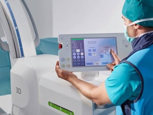
Service on a flat panel detector can be more complex than on an image intensifier, which seems like a disadvantage, but the point is moot if you opt to rely on an Original Equipment Manufacturer-certified service provider. It’s true that your technologists on site may be able to troubleshoot some issues inherent to image intensifiers, but this may not be the case with critical errors, which is why many facilities opt to go with a service contract. In these instances, the flat panel detector may make more sense. That way, the regularity of repairs can be reduced and, when repairs do need to occur, you receive care you know you can rely on.

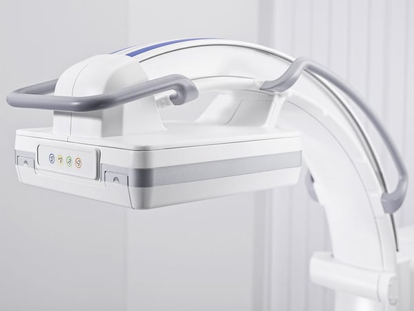
Size
This comparison is short (literally) and sweet. A flat panel detector is shorter than the image intensifier because, well, it’s a flat panel rather than an extended tube structure.
From a comfort standpoint, this leaves more room for your team members to navigate, because you’re gaining space between the patient and the technology. This is a definite positive whenever you’re operating on larger patients, as it enables you to maintain precise control without sacrificing ergonomic sensibility.
If, however, you’re handling simpler cases such as pain management rather than more complex procedures requiring precise movements and measurements, the size of the machine may not be a deciding factor.
Image Quality
There are two areas where flat panel technology wins out over image intensifiers: field of view and precision imaging.
Let’s start with field of view. For the comparison, I want you to think about a flat screen television versus a telescope. The flat screen provides a wide image that allows you to cover wide swaths of the patient’s anatomical structure as needed. The telescope can provide a fantastic image too, but you have to point it precisely or pull back sufficiently far in order to get the image you need within the viewer.
That’s a simplistic comparison, but I’ve found that it helps when trying to visualize the difference between the two. Flat panel technology can provide up to a 50% greater field of view than a similar class of image intensifier.
There’s also a difference when imaging smaller structures of the patient’s body. Flat panel detectors have a higher contrast resolution than image intensifiers, with the extra benefit of additional grayscale. These two things combine to provide precise views of anatomical structures that might appear out of focus using an image intensifier, especially if the latter system has been in commission for a few years and already experienced significant fatigue.
Cost
The final comparison I want to make between the two technologies is cost, and this one is also pretty straight-forward.
An image intensifier will most likely be the lower cost option in comparison with flat panel technology. The flat panel is newer, whereas image intensifiers have been around since the 1950s (a testament to both their longevity and their somewhat outdated technological foundation). Plus, flat panel detectors, as described above, tend to have better image quality thanks to field of view and precision imaging capabilities, and their development reflects the more recent evolution of technology.
When it comes to cost, image intensifiers will almost always cost less than flat panel detector technology. However, that cost doesn’t necessarily take into account the need for service (image intensifiers may experience increased downtime due to degradation and the need for parts replacement) or the fact that the technology that drives flat panel detectors has matured and become more readily accessible every year. As more options have been introduced, the cost of many flat panel detectors has come down considerably compared to even just a couple years ago.
The Right Choice Depends on Your Needs
Flat panel detectors and image intensifiers each have their strengths. It’s up to you to determine what your needs are and which system has the right qualifications for your facility.
Remember, too, that in addition to outright purchases, C-arm systems all come with a variety of implementation options. These include renting, leasing, financing and other creative means of securing the technology that will drive surgical volume and an effective patient and technologist experience at your organization.
To learn more about more flat panel detectors and image intensifiers, subscribe to our blog.


Comments