
 Advances in ultrasound technology, combined with hospitals looking to provide highly accurate yet affordable care to patients, have led to a shift in the way ultrasound is viewed. Exams are now being used to complement or even take the place of the types of diagnostic procedures that previously would have required an MRI, a CT, a biopsy and more.
Today, I’m going to look at why this is and provide insights into the types of procedures and diagnoses ultrasound is increasingly being used for.
The Sound of the Future
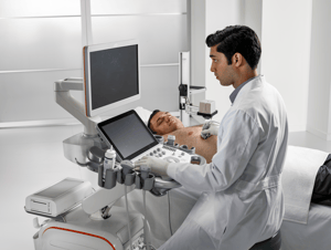
It’s easy to see the appeal of ultrasound. It’s less costly, less invasive and can provide for a better patient experience than other forms of testing. After all, you’re not asking your patients to submit to a painful, intrusive biopsy or telling them to climb into a large tube where they’ll have to remain prone for however long the examination takes. Instead, the sonographer uses a transducer to attain an accurate scan with relatively minimal discomfort.
Clearly, patients prefer ultrasound, but for a myriad of reasons, so do many administrators. The smaller footprint and increased mobility mean rooms can be converted to ultrasound suites on the fly as needs arise, with no need to worry about equipment transport or securing the room with lead-lined walls. Plus, the cost of an ultrasound is minimal compared to other types of diagnostic equipment.
The Centers for Medicare and Medicaid Services (CMS) are also coming around on reimbursable ultrasound procedures. As the Food and Drug Administration (FDA) approves more and more uses of the technology, CMS is following suit by issuing updated reimbursement rates for ultrasound procedures centered around the breast, the liver and more (additional info on these procedures further down).
 Tech Me Home Tonight
Of course one reason for the growth is the numerous improvements we’ve seen to ultrasound technology in recent years.
These range from ergonomic benefits that assist the sonographer performing the scan to diagnostic and workflow improvements that ensure reproducible, step-by-step imaging procedures. Meanwhile, ultrasounds are achieving new levels of accuracy, particularly with patient populations that have historically proven difficult to scan.
Let’s look at a few examples.
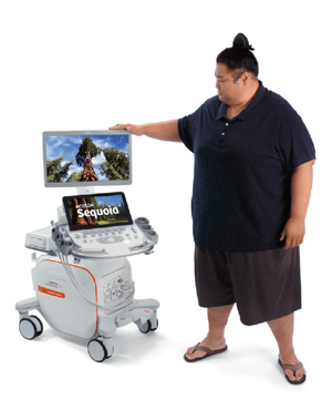
We’ll start with the ACUSON Sequoia. Launched about a month ago, the Sequoia was designed specifically for accurate, easy scanning of overweight patients. In fact, thanks to a technology developed by Siemens Healthineers called the Deep Abdominal Transducer, or DAX, it’s possible to achieve penetration of 40 cm. The goal is to improve diagnoses while simultaneously reducing rescans and ensuring image quality remains pristine.
 Another member of the ACUSON family of ultrasound systems, the Bonsai, was designed for cardiac ultrasound applications. This particular system was built for maneuverability and ease of use in mind. “Auto EF” creates semi-automatic ejection fraction measurements while “Auto IMT” enables automatic intima-media thickness measurements, the combination of the two resulting in one-click calculation. Combined with one-touch image optimization and enhanced portability, ACUSON Bonsai is an innovation that benefits both patients and providers.
It’s easy to see, even from just these two examples, why ultrasound is so appealing. Ultrasound is, dare I say, win-win-win. Providers, patients and payers, whether that payer be CMS or a private insurer, all prefer ultrasound procedures when possible, and that’s leading to more and more exams relying on this extremely convenient method of diagnostics.
Testing, Testing, 1,2…
Ultrasound is being used today in ways that may surprise traditionalists, but healthcare providers of all types are seeing the benefits and making inroads to clinical uses that were previously unheard of.
The use of contrast in ultrasound has, for example, completely altered the way a provider can approach the diagnosis of liver lesions. The standard used to be a plethora of tests that were harder on the patient and required additional touch points from multiple members of the care team.
Today, a single ultrasound exam can often take care of the procedure on its own. Through the use of contrast-enhanced ultrasound, it’s possible to monitor the filling pattern of a suspicious lesion to determine areas of further intervention and those areas that are benign. This keeps healthy patients from enduring an additional biopsy, and it can act as an alert for when at-risk patients should be routed to the additional exam or treatment they need.
Shear wave elastography is another type of ultrasound exam whose popularity has skyrocketed in recent years. By using force displacement, it’s possible to get an accurate representation of the state of abnormal tissue, deducing whether or not it’s hard or soft, allowing the physician to make their diagnosis based on these details. Currently, shear wave elastography is helping in two key areas: liver fibrosis and breast lesion stiffness.
 Breast density testing is something that’s becoming more and more common as well (and many states have passed laws related to breast density notification). That’s because density can be a key indicator of a woman’s risk of breast cancer. Unfortunately, the presence of dense tissue can make it difficult for a mammogram to detect the presence of cancerous growths.
This is problematic, considering that almost 50% of women may have extremely radiographically dense breast tissue. This is why Automated Breast Volume Scanners (ABVS) like the ACUSON S2000 are so important.
The S2000 provides 3D Total Breast Ultrasound in order to gain a more accurate assessment of abnormalities within dense breast tissue. By generating a coronal view of the breast, it’s possible to see global anatomy and symmetry and thus identify potential areas of diagnostic interest. Tissue stiffness analysis via shear wave elastography allows the diagnostician to categorize suspected lesions in a non-invasive manner, making it possible to identify cysts, tumors and more.
Ultrasound is also having an impact on coronary procedures, leading to less invasive techniques that increase the patient’s chances of survival and recovery. Because ultrasound can be used to guide the placement of a stent in individuals experiencing cardiac trauma, the need for open heart surgery and all its attendant risks is far less than it was a decade ago. Cardiac ultrasound for emergency procedures is yet another way ultrasound is supplanting other tools in the collective conscious of the healthcare world.
It’s an Ultrasound World…We’re Just Living In It
Let’s be clear: hospitals aren’t getting rid of their CT or MRI systems anytime soon. Certain procedures will always require a degree of calculation and visibility that ultrasound waves aren’t able to provide.
But more and more, the dividing lines between operations and exams using one type of technology over another have blurred. This is no different to the blurring of lines between members of a care team, with healthcare providers of all types seeing themselves as parts of a single unit that has the patient’s best interest at heart.
Ultrasound will only cement its place in healthcare further in the coming years. It may not ever be as ubiquitous as the stethoscope, but it could get pretty darn close.


Comments